
The latissimus dorsi muscle is a broad, flat muscle that occupies the majority of the lower posterior thorax.
The latissimus dorsi is a large, fan-shaped muscle located on the back and is one of the superficial extrinsic back muscles. In this blog post, we will explore the anatomy of the latissimus dorsi, including its origins, insertions, innervation, blood supply, and functions.
Functions
The latissimus dorsi has attachments on both the axial and appendicular skeletons, so it produces movements for both the trunk and the upper limb.
The main functions of the latissimus dorsi are extension, adduction, and medial rotation of the humerus. It works with the teres major and pectoralis major to produce adduction and medial rotation, and with the sternal head of the pectoralis major and teres major for extension.
It also helps bring the body towards the arms when the upper extremities are fixed overhead, such as in climbing or during chin-ups. Additionally, the latissimus dorsi can serve as an accessory muscle of respiration and has been found to be active during deep inspiration and forceful exhalation, such as when sneezing or coughing.
Anatomy

The majority of the lower posterior thorax is covered by the large, flat latissimus dorsi muscle. Although the muscle’s main use is for the upper extremities, it is also regarded as a respiratory accessory muscle (1).
Origin
The lower six thoracic vertebrae on the posterior layer thoracolumbar fascia, supraspinous ligaments of all lumbar vertebrae, sacral vertebrae and posterior crest of ilium, lower 3 or 4 ribs, and tip of the scapula (2).
Insertion
After the fibers twist around each other, they insert on the humerus’ intertubercular groove just anterior and parallel to the pectoralis major tendon (2).

The muscle fibers converge superolaterally towards the axilla, winding around the anterior aspect of the teres major muscle, before inserting via a flat tendon onto the anterior aspect of the proximal humerus on the floor of the intertubercular sulcus.
Action
Adduct and medially rotate the humerus with teres major and pectoralis major, extend the humerus with teres major and the sternal head of pectoralis major, and move the trunk forward and upward in activities such as climbing or chin-up.
Respiratory functions include coughing, sneezing, and deep inspiration (1).
Innervation
The innervation of the muscle is by a branch of the posterior cord of the brachial plexus, the thoracodorsal nerve through the spinal nerves C6 to C8 with C7 predominant (1)(3).
The muscle is predominantly supplied by the thoracodorsal branch of the subscapular artery, which is the largest branch of the axillary artery arising from its third part.
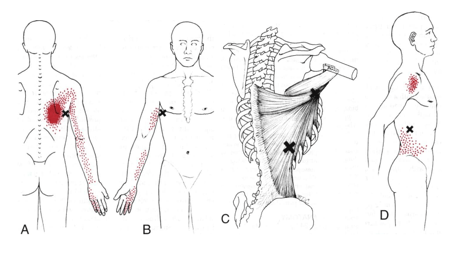
Pain Pattern
Usually, referred pain from trigger points in the muscle is readily misunderstood as emanating from intrathoracic abnormalities (3).
Pain is located in a 5-10 m zone at the lower angle of scapula which radiates to the dorsal shoulder area and to the medial aspect of the arm, forearm, and ulnar aspect of hand affecting fingers 4 and 5 (4,5).
Triangular pattern from lower lateral trigger point radiates to the brim of the pelvis (5).
Trigger points
“A myofascial trigger point is defined as a hyperirritable spot in skeletal muscle, which is associated with a hypersensitive palpable nodule in a taut band.”(6)
Pincer palpation of the latissimus dorsi in the posterior axillary fold at roughly the midscapular level with the arm in lateral rotation and abducted by about 90 degrees to fill up the slack is required for a trigger point examination. At the midscapular level, where the latissimus dorsi wraps around the teres major, the examiner grasps the latissimus dorsi muscle along the free border of the posterior axillary fold.
The firm bands and their points of greatest tenderness are rolled between the fingers and thumb as the muscle is raised off the chest wall. Typically, the apex of the arch of the posterior axillary fold is a few centimeters (or approximately an inch) below where these TrPs are located. Depending on which fibers are implicated, palpating one of these bands causes a significant local twitch response that can be easily observed along the scapular border or over the lower thoracic and lumbar areas.(3)
Careful examination of tasks that demand strong shoulder-girdle depression (weight bearing) or repetitive extension, particularly when accompanied with adduction, is necessary to pinpoint the cause of insidious activation. Instead of when the muscle’s depressor and arm extension functions are overloaded, symptoms are more likely to be felt when it is stretched by reaching up and forward (3).
Reaching too far to exercise by lifting heavy weights, throwing a baseball, hanging from a swing or rope, and pressing down to twist weeds out of the way when gardening are a few common movements that can eventually activate these TrPs (3).
Satellite trigger points for the latissimus dorsi includes the serratus posterior superior muscle, teres major, triceps brachii, rectus abdominis, iliocostalis thoracis, and iliocostalis lumborum (4).
With the body pressure to a latissimus TrP while sleeping, pain can be activated with the risk of substantially disrupting the sleep and the ability to function the following day.
Once triggered, moving the arm to help get up or down from a low seat will exacerbate TrPs in the latissimus fibers that are more vertical (3).
Exercises for Latissimus Dorsi Pain
In this comprehensive guide, we will explore a step-by-step process to release, lengthen, and strengthen these muscles, potentially providing relief within 30 seconds.
These exercises can be done at home and are designed to work together for the best results.
Part 1: Warm-up and Muscle Tightness Release
To prevent muscle irritation and improve circulation and flexibility, it’s essential to warm up the muscles and soft tissue before exercising. You can use a tennis ball or a rolled-up sock for this exercise.
- Stand next to a wall with your painful side facing it and your arm raised upward.
- Lean your body slightly away from the wall, placing the tennis ball or sock between the wall and your lat muscle.
- Apply comfortable pressure and glide the ball over the muscle for about 15-20 seconds, focusing on tight spots.
- Experiment with different movements, like going up and down, forward and backward, or in circular motions.
- If you find a particularly tight spot, press into the ball and rotate your upper body to open up the muscle further.
Part 2: Strengthening Exercises
To achieve long-lasting relief, it’s crucial to strengthen your lat muscles. Here is a daily strengthening exercise that you can do in bed or on the floor:
- Lie on your stomach with your shoulders and head off the edge of the bed.
- Perform three different motions (alternating bring back, baby penguin, and snow angel) to target the lat muscles.
- Do each exercise slowly for 10-15 repetitions, gradually increasing intensity.
Part 3: Stretching
Stretching is important after exercising to keep muscles loose and prevent soreness. Try these stretches for quick pain or tightness relief:
- Door Frame Stretch
- Stand next to a door frame and grip overhead with your arm straight.
- Lean away from the frame and rotate your hips towards it.
- Lean away again to deepen the stretch, holding for 20-30 seconds.
- Modified Child’s Pose
- Begin on all fours on the floor, with a pillow or towel under your knees for comfort.
- Reach your painful side’s arm across your body, bending the upper body and moving through the upper back and shoulder blades.
- Lower your chin to your chest and lower your buttocks towards your heels.
- Hold the stretch for 20-30 seconds, repeating three to five times on each side.
References
- Varacallo M. Anatomy , Back , Latissimus Dorsi Anatomy , Back , Latissimus Dorsi. 2018;(December):1–4.
- Bhatt CR, Prajapati B, Patil DS, Patel VD, Singh BGP, Mehta CD. Variation in the insertion of the latissimus dorsi & its clinical importance. J Orthop [Internet]. 2013;10(1):25–8. Available from: http://dx.doi.org/10.1016/j.jor.2013.01.002
- DAVID G. SIMONS, JANET G. TRAVELL LSS. Travell & Simons’ myofascial pain and dysfunction – the trigger point manual Volume 1. 2nd editio. 1999. 1052 p.
- Practice T, Touch I, Arts H, Rochester P. Trigger Point Therapy for Myofascial Pain. 2005. 311 p.
- Niel-asher S. The Concise Book of Trigger Points. 2nd editio. 2008. 217 p.
- Donatelli RA. Physical therapy of the shoulder. 5th editio. 2012. 489 p.
MD, PhD. Physical Medicine & Rehabilitation Physician from São Paulo - Brazil. Pain Fellowship in University of São Paulo.